Implant Failures
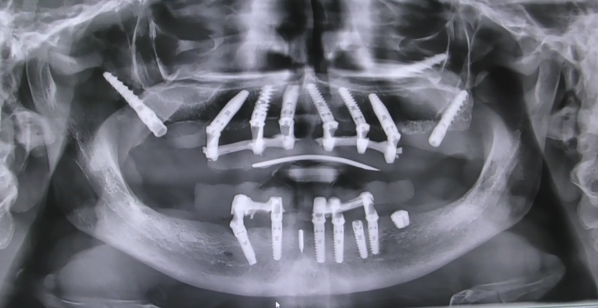
Causes of failure
The main causes of implant failure are peri-implantitis (~40%), an infectious inflammation that destroys the peri-implant alveolar bone, occlusal overload (~25%) that causes micro-fractures and resorption, implant fracture (~15%) and other causes such as failed osseointegration, positioning errors and systemic causes.
The main causes of dental implant failure, with their approximate incidence:
| Cause | Incidence | Description |
|---|---|---|
| Peri-implantitis | ~40% | Inflammation and infection of the tissues around the implant, with progressive loss of alveolar bone. It is the most common cause of late failure (>1 year from loading). Related to poor oral hygiene, smoking, previous periodontitis and poorly adapted prosthesis |
| Occlusal overload | ~25% | The masticatory forces exceed the load capacity of the implant or the surrounding bone. It causes micro-fractures of the peri-implant bone, resorption and loss of osseointegration. More common in the posterior sectors and in patients with bruxism |
| Implant fracture | ~15% | Fracture of the implant body, of the abutment screw or of the prosthetic structure. Caused by overload, manufacturing defects, reduced implant diameter or inadequate alloys |
| Other causes | ~20% | Failed initial osseointegration, positioning error, nerve damage, chronic sinusitis, systemic causes (uncontrolled diabetes, immunosuppression, bisphosphonate therapies) |
For a more detailed treatment of the causes and risk factors, visit the Risks and Complications page.
Diagnosis
The early diagnosis of implant failure is based on clinical signs such as bleeding on probing, suppuration, mobility and pain on percussion, on radiographs showing peri-implant bone loss above 50%, clinically detectable mobility indicating irreversible failure and Cone Beam CT to evaluate the residual bone for a retreatment.
The early diagnosis of an implant at risk of failure is based on:
- Clinical signs: bleeding on probing, suppuration, mobility, pain on percussion, gingival recession, persistent halitosis
- Radiographs: visible peri-implant bone loss on intraoral radiographs (parallel technique) or on orthopantomograms. A bone loss >50% of the implant length is an indicator of imminent failure
- Mobility: every implant with clinically detectable mobility must be considered failed and removed, because the osseointegration is irreversibly compromised
- Cone Beam CT: to precisely evaluate the extent of the bone loss and the morphology of the residual bone for a possible retreatment
Prevention
The prevention of implant failures is based on correct planning with adequate number and diameter of implants, scrupulous daily oral hygiene with toothbrush and floss, periodic checks every 6-12 months with a hygienist and annual radiograph, night bite for patients with bruxism and reduction of risk factors such as smoking and diabetes.
- Correct planning: adequate number of implants, appropriate diameter and length, optimal biomechanical distribution
- Oral hygiene: scrupulous daily hygiene with toothbrush and floss, especially around the prosthesis
- Periodic checks: every 6-12 months with professional hygiene and annual radiograph
- Night bite: for patients with bruxism, to protect the implants from nocturnal overload
- Risk factors: smoking cessation, diabetes control, treatment of parafunctions
Removal and retreatment
In case of implant failure, the implant is removed with counter-torque or trephine, the socket is cleaned and decontaminated, and after healing of 2-4 months a new implant can be placed or an alternative technique such as the Bilateral Sinus Solution™ (BSS) can be used.
When an implant fails, the procedure is:
- Removal of the failed implant: with counter-torque or trephine
- Cleaning and decontamination of the socket
- Healing of 2-4 months
- Retreatment: placement of a new implant or alternative technique such as the Bilateral Sinus Solution™ (BSS)
Visit the page Failed Implant Removal for details.
How BSS prevents failures
The Bilateral Sinus Solution™ (BSS) prevents failures thanks to the anchorage in the D1-D2 basal bone that does not resorb, to the absence of micro-movements thanks to the bicortical anchorage, to the prosthesis without fake gum that facilitates hygiene and prevents peri-implantitis, to the posterior molars that distribute the loads and to the elimination of cantilevers and inclined implants.
The Bilateral Sinus Solution™ (BSS) prevents failures thanks to:
- Anchorage in the D1-D2 basal bone that never resorbs
- Absence of micro-movements thanks to the bicortical anchorage
- Prosthesis without fake gum that facilitates hygiene and prevents peri-implantitis
- Posterior molars that distribute the loads over the whole arch
- Elimination of cantilevers and angled implants
Discover how the BSS solves all the failures of the traditional techniques.
The Circolare d'Autore® prosthesis
- 14 teeth per arch, including all posterior molars
- Zero fake gum: the teeth emerge from the natural gum
- Zirconia + layered ceramic, with registered trademark ®
- Custom-made: every prosthesis is a unique piece designed for the patient
Discover all the details on the dedicated page: Circolare d'Autore® — the definitive prosthesis without fake gum.